
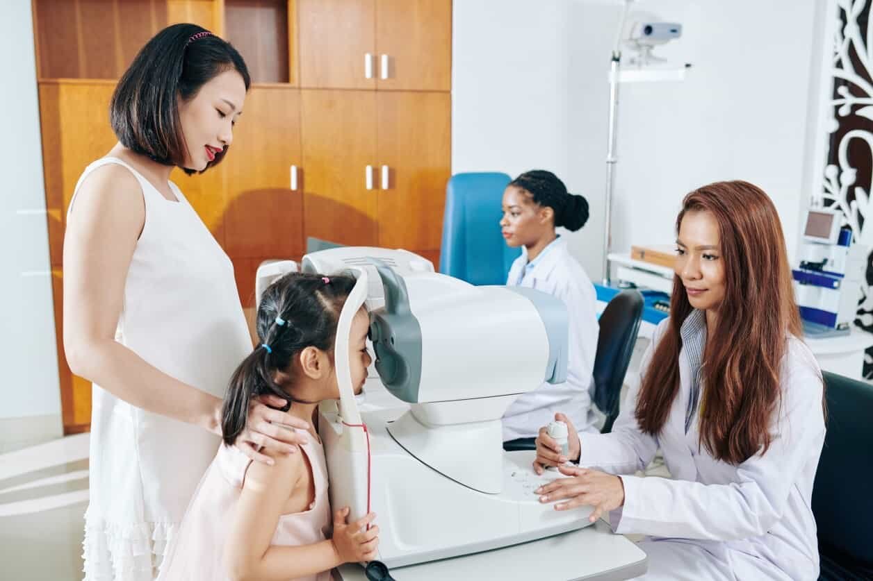
Fundus cameras are essential for optometrists and ophthalmologists in today’s rapidly advancing eye care landscape. These special imaging devices allow clear views of the retina, optic nerve, macula, and back of the eye. They help eye care professionals accurately find, track, and record eye and body diseases. The right camera serves as the “eyes” of your practice.
The demand for digital retinal imaging is increasing. This is because of more cases of diabetic retinopathy, age-related macular degeneration (AMD), and glaucoma. As people get older and chronic diseases increase, early detection with good imaging is more important than ever.
Technological innovations have changed fundus cameras. They are no longer just basic diagnostic tools.
Now, they are key parts of complete eye care. They support telemedicine, automated screenings, and AI-powered analysis. They also work well with practice management software.
In short, the right technology isn’t just a “nice to have.” Fundus cameras improve optometry skills, help diagnose important conditions early, and let optometrists expand their practice and better serve their community.
Let’s explore the value of a fundus camera for your practice. We will also review the 2025 buyer’s guide for optometry professionals.
What Is a Fundus Camera and Why Is It Essential?
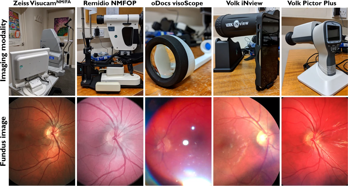
A fundus camera is a special low-power microscope. It has a camera that takes pictures of the inside of the eye. It provides clear, detailed images of the retina, optic disc, macula, and posterior pole. These images are invaluable in diagnosing, monitoring, and managing ocular and systemic diseases.
Why fundus cameras are indispensable in modern eye clinics:
- Early disease detection: Fundus photography helps doctors find early signs of diabetic retinopathy, AMD, glaucoma, and hypertensive retinopathy. This can happen years before any symptoms appear.
- Documentation: High-quality images provide a baseline for future comparison, facilitating longitudinal monitoring of disease progression and treatment efficacy.
- Telemedicine allows healthcare providers to securely share fundus images with remote specialists, supporting collaborative care and improving access in underserved areas.
- Workflow improvement: Automation and EMR integration reduce manual data entry, reducing errors and allowing clinical staff to focus more on patient care.
- Patient education: Visual evidence of ocular health status helps patients better understand their conditions, promoting compliance with treatment plans.
The fundus camera gives providers and patients a clear look into their ocular health. This builds trust, confidence, and rapport while elevating diagnostics and streamlining workflows. Investing in a high-quality camera is one the best choices a practice manager can make for their patients.
Key Criteria for Choosing the Best Fundus Camera
Choosing the right fundus camera can be tough. However, focusing on key criteria will help you make a good choice.
Image Resolution and Field of View
High-resolution images are essential for detecting subtle pathologies. Most cameras offer a field of view between 30° and 60°, though ultra-widefield models can capture up to 200°. A larger field reduces the need for multiple images and can improve diagnostic efficiency.
Mydriatic vs. Non-Mydriatic Operation
- Mydriatic cameras require pharmacological pupil dilation. They often produce superior image clarity, especially in challenging cases.
- Non-mydriatic cameras allow imaging without dilation, increasing comfort and speed, and are ideal for high-volume clinics.
Some devices offer both modes, providing greater flexibility. Consider whether a dual-mode option is right for your practice, depending on your budget and needs.
Ease of Integration with EMR Systems
Any practice manager knows that when EMR systems work well with technology, the team saves time and stress. This may be one of the most important elements when upgrading your optometry tech.
Your fundus camera should work well with your EMR system, DICOM networks, and practice management software. This will help ensure a smooth workflow, keep data secure, and meet regulations. Be sure to test the camera out before committing to the purchase.

Portability and Workflow
Are you a provider on the go? Do you and your team often travel to schools, rural communities, and nursing homes?
What about community support efforts? A compact or portable model may be essential for offering outreach programs, mobile services, or multiple locations. Consider whether you can easily relocate the camera or use it handheld for added versatility.
Budget and After-Sales Support
Look beyond the sticker price to understand the full lifetime value of new equipment. Be sure to factor in service agreements, warranty coverage, software updates, training, and technical support. A lower upfront cost may not translate into better long-term value.
Additional Considerations
- AI compatibility: Can the device integrate with AI-assisted diagnostic platforms?
- User training: Is the camera intuitive for staff with varying experience levels?
- Ergonomics: Is the device comfortable and easy for the patient and the technician?
The Top 5 Fundus Cameras Used by Optometrists in 2025
Get a clear visual on the latest technology for optometrists in 2025. These are the top 5 fundus cameras optometrists are using today.
1. Topcon TRC-NW400
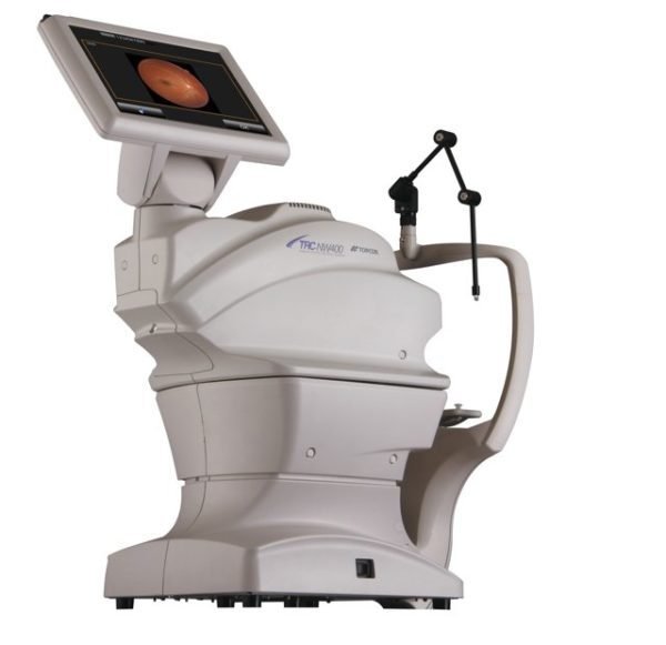
Features:
- 45° non-mydriatic field of view
- Fully automated alignment, focus, and capture
- Touchscreen interface
- DICOM-ready for EMR integration
Pros:
✅ User-friendly for clinics of all sizes
✅ Consistent, high-quality imaging without dilation
✅ Small footprint
Cons:
❌ Higher cost compared to basic models
❌ Not suitable for ultra-widefield imaging
2. Canon CR-2 AF Digital Non-Mydriatic Fundus Camera
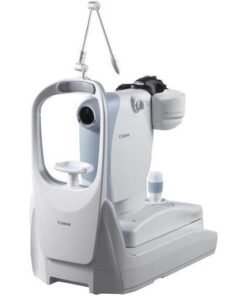
Features:
- 45° field of view
- Auto-focus and auto-shoot functions
- Low flash intensity for enhanced patient comfort
- Compact design
Pros:
✅ Excellent for routine screenings
✅ Minimal training required
✅ Low maintenance
Cons:
❌ No widefield imaging capability
❌ Lacks portability
3. ZEISS CLARUS 500 Fundus Camera

Features:
- True color ultra-widefield imaging up to 200°
- Mydriatic and non-mydriatic modes
- Auto-montage for composite imaging
- Advanced DICOM/EMR compatibility
Pros:
✅ Unmatched image quality and color fidelity
✅ Captures peripheral retina with minimal images
✅ Ideal for complex pathology documentation
Cons:
❌ High investment cost
❌ Larger footprint
4. Nidek AFC-330 Automated Fundus Camera
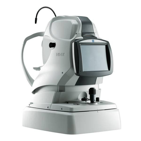
Features:
- Non-mydriatic 45° field of view
- Fully automated alignment, focus, and shooting
- Compact design for small exam rooms
Pros:
✅ Highly efficient for high-volume practices
✅ Easy for technicians to use
✅ Space-saving
Cons:
❌ Limited FOV
❌ Not portable
5. Optomed Aurora IQ Portable Fundus Camera

Features:
- Handheld non-mydriatic design
- 50° field of view
- Wireless image transfer
- Rechargeable battery
Pros:
✅ Ideal for mobile screenings and telemedicine
✅ Lightweight and versatile
✅ Lower upfront cost
Cons:
❌ May not match tabletop units in image detail
❌ Limited battery life
Do you need a little more help deciding which camera is optimal for you? We’ll help you prioritize your practice’s needs based on which features to remember during your buying journey. Read on for simple tips on choosing the right fundus camera.
Expert Tips: How to Pick the Right Fundus Camera for Your Practice
When deciding on a fundus camera, keep these expert insights in mind:
- Match technology to practice type: A community clinic might need a portable solution. A specialist center may focus on ultra-widefield imaging.
- Future-proof your investment: Look for devices with upgradable software, AI integration potential, and strong manufacturer support.
- Test before you buy: Whenever you can, set up a demo. This will help you check if it is easy to use and if the image quality meets your needs.
- Evaluate long-term costs: Consider consumables, service contracts, and software licensing fees.
Considering these guiding tips, choosing a fundus camera that scales with your practice will benefit your team in the long term.
FAQs: Fundus Cameras for Optometry Clinics
Purchasing new equipment is a big decision. If you still have lingering questions, chances are we’ve heard them before. Here are some of the most asked questions from our optometry community.
What’s the difference between mydriatic and non-mydriatic cameras?
Mydriatic cameras require dilation, offering superior image quality in difficult cases or with small pupils. Non-mydriatic models capture images without dilation, providing faster exams and greater comfort.
How often should I calibrate my fundus camera?
Experts typically recommend annual calibration, but high-volume practices or those in research settings may require more frequent checks.
Are portable fundus cameras reliable for routine practice?
Yes — today’s portable fundus cameras provide high-quality images suitable for screenings, outreach, and supplemental use in clinics.
What is ultra-widefield imaging, and do I need it?
Ultra-widefield imaging captures up to 200° of the retina in one image. This helps document problems in the outer areas better. It’s beneficial for diabetic retinopathy, retinal tears, and uveitis cases.
Can fundus cameras integrate with AI platforms?
Many modern fundus cameras are compatible with AI software, enhancing diagnostic capabilities by automating the screening of diabetic retinopathy, AMD, and glaucoma suspects.
If you don’t see the answer to your question here, you can contact our team of experts. Dauh Eye Care is here to support your practice’s growth and success.
Why Optometrists Choose Dauh Eye Care
Dauh Eye Care is more than a supplier. We’re your strategic partner in advancing optometry. As a high-tech expert in eye equipment, we are proud to provide complete solutions to clinics and hospitals in more than 60 countries.

They founded Dauh Eye Care to bring together new ideas and strong support. They create, distribute, and support many types of optical diagnostic equipment. Our carefully curated lineup includes fundus cameras, OCT systems, slit lamps, auto-refractor/keratometers, phoropters, corneal topographers, and more.
At Dauh Eye Care, we do more than sell products—we deliver solutions. We select each device in our catalog for three main reasons. First, it has strong clinical performance.
Second, it has an easy design and works well with modern EMR systems. Third, we provide reliable equipment for your needs, whether you run a mobile screening service or a busy specialty practice.
Optometrists across the country trust Dauh Eye Care for their fundus imaging needs. They have a strong relationship and a history of quality and trust.
- You can access top brands and the latest models. Whether you need Topcon, ZEISS, Canon, Nidek, or Optomed, Dauh Eye Care has a wide selection for your practice.
- Award-winning customer support: Our team is with you every step from the first meeting to after-sales training. We offer support for questions, concerns, and any inquiries or issue that may arise. We can even guide you in selecting the best fundus camera for your practice’s needs and future goals.
- Flexible financing: We provide customizable financing options to help you manage your investment wisely. We know running a practice is costly so let us help make the financing process stress-free and affordable.
- Real-world success: Our clients consistently praise our responsiveness, expertise, and commitment to their success. Being a part of our client community means having a supportive partner throughout your practice’s journey.
Discover the difference of excellence when choosing Dauh Eye Care for all your equipment needs.
What’s Next? Choosing The Right Fundus Camera for Your Optometry Practice
Ready to move forward with your practice’s setup and equipment? If you feel confident and informed by this simple guide, our team will help you take those next steps.
The right fundus camera isn’t just a device — it’s a critical partner in delivering high-quality eye care. Whether focused on early disease detection, telemedicine, or streamlining your workflow, the ideal camera should meet your clinical needs today while adapting to future advancements.
Check out Dauh Eye Care’s fundus cameras for optometrists. You can also schedule a demo to see how the right imaging solution can improve your practice.