


Zeiss Clarus 700 Ultra widefield Fundus Camera enables doctors to detect even the smallest changes in the course of the disease. In addition to true-color fundus images, the system also captures high-resolution fundus autofluorescence (FAF) images in FAF blue and FAF green, infrared images (IR), and external images. The ultra-high resolution of the CLARUS 700, combined with our intuitive review software, enables clinicians to track changes confidently.
Zeiss Clarus 700 was designed as a comprehensive ultra-widefield retinal camera for eye care specialists to capture ultra-widefield images in true color, with unsurpassed image quality and a complete suite of modalities including fluorescein angiography.
Key Features:
- Ultra-wide field of view: Captures images from the macula to the far periphery, up to 200° with single capture and 267° with montaging.
- True Color Imaging: Uses a combination of red, green, and blue LEDs to produce images that closely resemble what is seen during a clinical exam.
- High Resolution: Provides sharp, detailed images from the posterior pole to the periphery.
- Fluorescein Angiography (FA): Enables detailed visualization of blood vessels and potential leakage in the retina.
- Fundus Auto-fluorescence (FAF): allows imaging with both blue and green excitation light.
- AutoBright: Automatically optimizes the brightness of an entire series of images for consistent exposure.
- PrecisionFocus: Allows quick focus optimization on a specific area of interest without losing focus on other areas.
- GazePoint: Uses AI to automatically identify the optic nerve and derive the patient’s gaze angle, improving efficiency and accuracy.
- Integration with ZEISS Integrated Diagnostic Imaging: Facilitates streamlined workflow and data management.
- Live Infrared (IR) Preview: Helps in patient positioning and alignment.
- QuickCompare: allows easy comparison of images over time and between different imaging modes.
- Ease of Use: Features like the chin rest that moves with the camera (not the patient) enhance patient comfort and image quality.
Benefits:
- Enhanced Diagnostic Precision: The combination of ultra-widefield imaging and high resolution allows for earlier and more accurate detection of retinal diseases.
- Improved Workflow: Features like AutoBright and GazePoint streamline the imaging process and reduce chair time.
- Better Patient Experience: Features like the chin rest and live IR preview contribute to a more comfortable and efficient examination.
- Comprehensive Imaging: The wide range of imaging modalities allows for a thorough assessment of various retinal conditions.
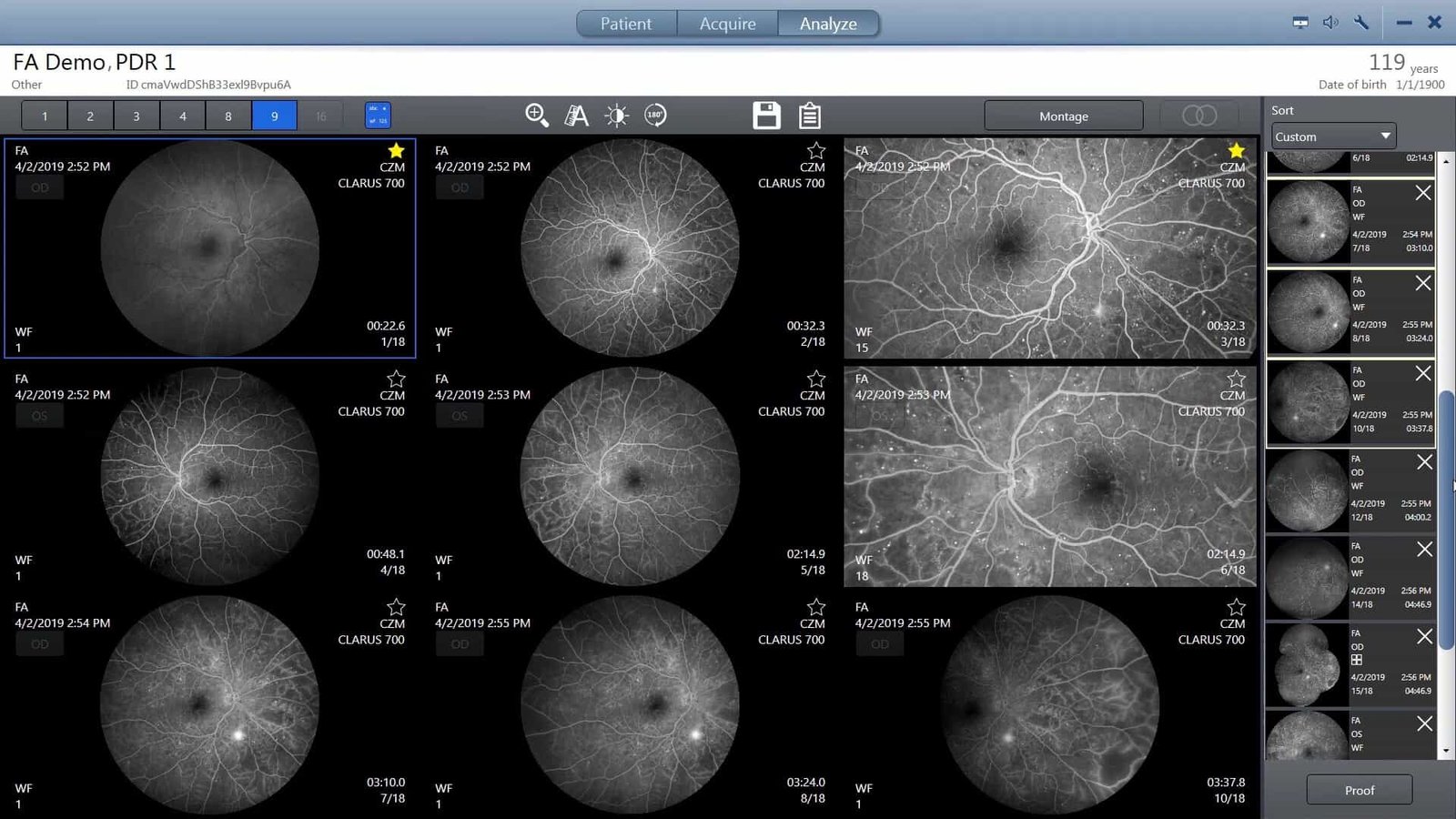
The Zeiss Clarus 700 captures images that closely resemble the coloration of the fundus as it appears under direct clinical observation. Ultra-widefield fluorescein angiography is a useful exam to identify diabetic retinopathy, especially visualizing the peripheral retina, which is fundamental to assessing nonperfused areas, vascular leakage, microvascular abnormalities, and neovascularizations.
Zeiss Clarus 700 enables you to capture sharp, precise images from the macula to the far periphery, all with a single instrument that combines:
- • Ultra-large field of view
• True color images thanks to wide spectrum LED scans
• Exceptional image resolution
• Fluorescein angiography
• Advanced image capture features