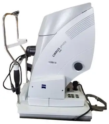
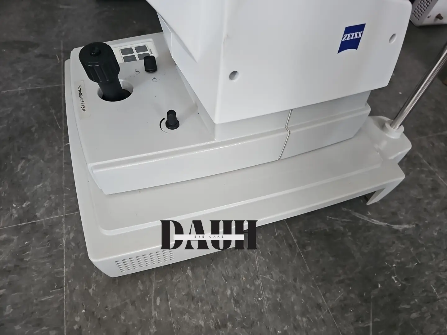


Zeiss Cirrus Photo 600 Optical Coherence Tomography combines a full mydriatic/non-mydriatic color camera and CIRRUS OCT technology in a single integrated and compact system with a fundus autofluorescence option that is designed for comprehensive eye care practices.
Zeiss and its research collaborators have developed advanced algorithms to measure and display layers. FoveaFinder™ and AutoCenter™ automatically ensure that measurements are made in the correct locations, taking the pressure off the operator to perfectly center the scans. CIRRUS data cubes are automatically registered with data from prior visits, allowing for more detailed comparisons.
Diversified normative databases for ONH, RNFL and macular thickness facilitate even more at-a-glance assessments. Enabling efficient cross-modality analysis, CIRRUS photo allows easy switching between fundus images registered with OCT scans and maps.
Zeiss Cirrus Photo 600 Key Features
- Multimode Navigator- interactive display of fundus image and OCT scans
- Precise registration and display of OCT scans with color fundus image
- Macular Thickness and Change Analysis
- Macular Thickness Normative Data
- ONH & RNFL OU Analysis with normative data
- Guided Progession Analysis
Why should you choose CIRRUS OCT Photo 600?
- Visualized findings from various modalities
- Correlated data from high-density OCT cubes
- Superb color fundus results
- Fundus autofluorescence and fluorescein angiography
- A more comprehensive clinical evaluation
- Very comfortable
- A non-invasive procedure
- Resume normal activities
Broader Clinical Insights – Greater Diagnostic Certainty
The Smart Imaging Combo
CIRRUS photo 600 combines a full mydriatic/non-mydriatic true color camera and CIRRUS OCT technology in a single integrated and compact system. Fundus autofluorescence is optional. CIRRUS photo 600 was designed with comprehensive eye care practices in mind. Broader clinical insights, greater diagnostic certainty and added practice value – the new Zeiss Cirrus Photo 600 from ZEISS delivers all that in a single, integrated system for both fundus imaging and OCT. All in one convenient sitting for our patints. Achieves a more comprehensive clinical evaluation. Comprehensive, high-quality diagnostics form the basis for informed decisions. With its superb multimodality visualizations, CIRRUS photo delivers exceptional diagnostic accuracy and certainty.
Interactive Review
The system’s one-of-a-kind Multi-Mode Navigator enables interactive analysis of registered fundus images and OCT cube scans – horizontal and vertical direction.
Precise Registration
OCT scans are automatically registered with different types of fundus images including color fundus, angiography and fundus autofluorescence* images, bringing depth to our analysis.
Multimodal Assessments
CIRRUS photo allows us to conduct examinations with various modalities and to correlate the findings at one single workstation. Every fundus image can also be registered independently of the acquisition sequence, along with other flexible combinations.
Orientation At A Glance
Whether for a quick overview or point-by-point comparisons, thumbnails provide at-a-glance insights.