Carl Zeiss Clarus 500 Ultra widefield Retinal Camera is the very first fundus imaging system that combines true color with exceptional clarity in an ultra-widefield view. “Early signs of eye disease can frequently be subtle and occur in the far periphery of the retina,” says Jim Mazzo, international president of ophthalmic apparatus at Carl Zeiss Meditec. “With this system, professionals can obtain a better perspective of the entire fundus.”
The system produces a 133-degree field of view with one catch, and its own high profile ultra-widefield graphics are automatically merged to achieve a 200-degree field of view.
Carl Zeiss Clarus 500 Ultra widefield Retinal Camera allows clinicians to review and compare high-quality pictures recorded during one exam, while also providing annotation and caliper measurement tools which allow for comprehensive evaluation of eye health, Zeiss said.
Carl Zeiss Clarus 500 Ultra widefield Retinal Camera is the first and only fundus imaging method to provide true color and clarity inside an ultra-wide field of view, allowing clinicians to catch high-resolution fundus pictures from macula into the far periphery.
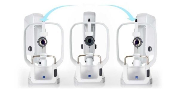
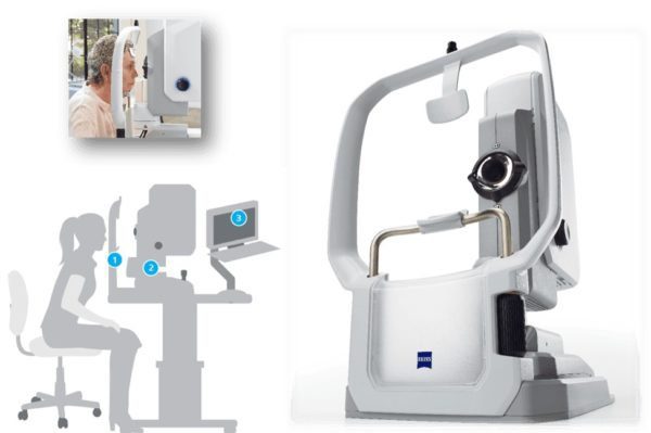
The Carl Zeiss Clarus 500 Ultra widefield Retinal Camera uses three wide-spectrum LEDs to enable image capturing in true color and cut back optic nerve head whitening. The partly confocal optics reduce lid, lash, and other artifacts from the anterior segment. External ocular images can also be obtained to aid in anterior segment disease management and instruction. The combines numerous technical features into a sleekly designed platform with both patient and operator comfort in mind
Carl Zeiss Clarus 500 Ultra widefield Retinal Camera Owing to its vast collection of technical features and its ease of use, the Carl Zeiss Clarus 500 Ultra widefield Retinal Camera can effectively function as only fundus camera in any office, irrespective of if that office is a large volume multi-specialty practice or a one-doctor clinic.

When reviewing numerous pictures, the Carl Zeiss Clarus 500 Ultra widefield Retinal Camera picture review software can coordinate the pictures to the nevus using a common zoom element.
The CLARUS may image peripheral hemorrhaging but also supply high-quality pictures of the macula. These images can be zoomed in to value the fovea for smaller microaneurysms.
Because of its broad scan size, The Carl Zeiss Clarus 500 Ultra widefield Retinal Camera may visualize other lesions inside the same eye or inside the fellow eye which are difficult to visualize with DFE or color photography. Disseminated or bilateral retinal findings may change not merely the prognosis but follow up and management of these patients.
With CLARUS 500, the latest in ultra-widefield retinal imaging, reveal more without compromise and manage your patients with confidence.
- See true color images to help with differential diagnosis and documentation
- Image in high resolution anywhere in the retina, including the periphery
- Elevate patient experience while driving clinical efficiency