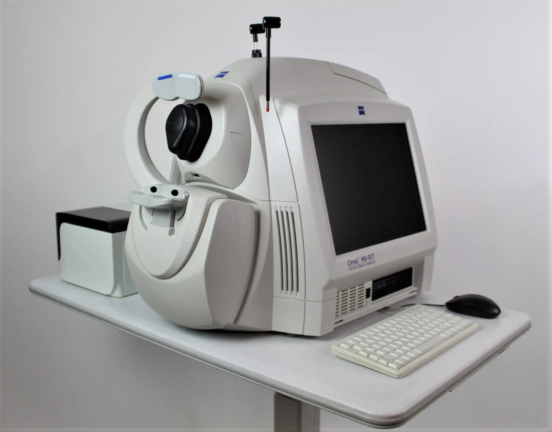
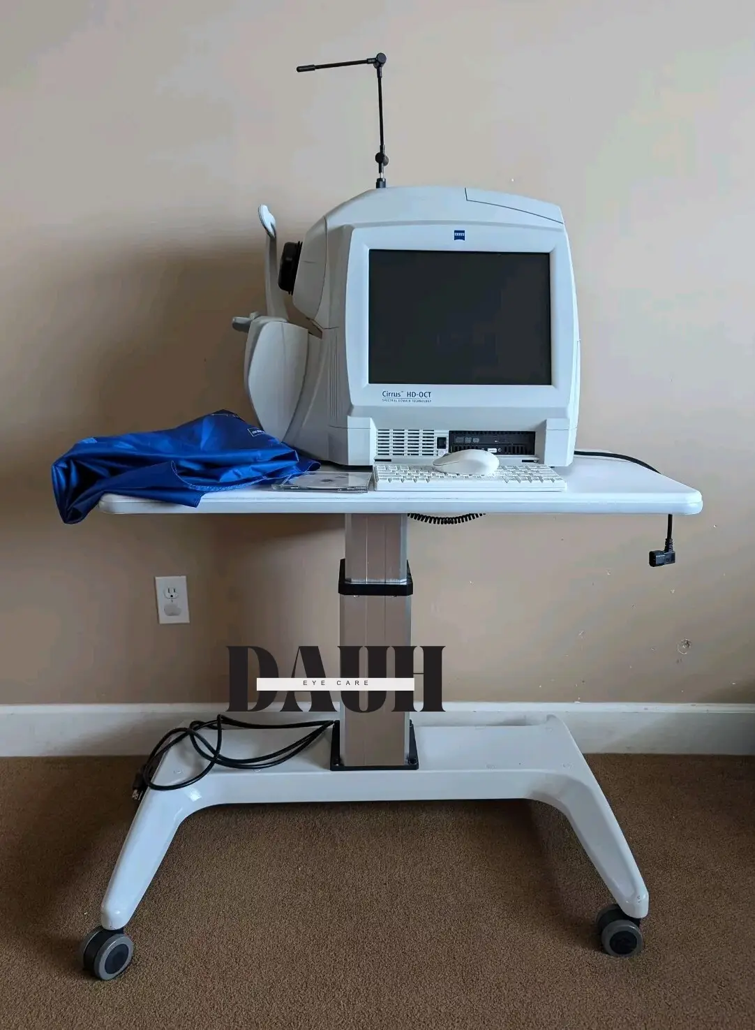
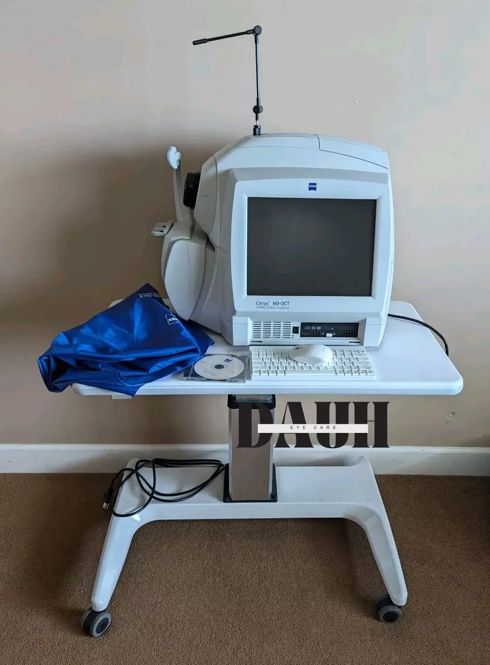
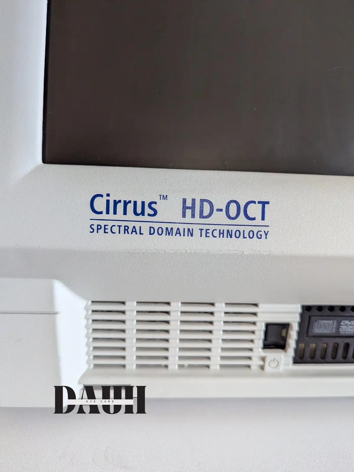
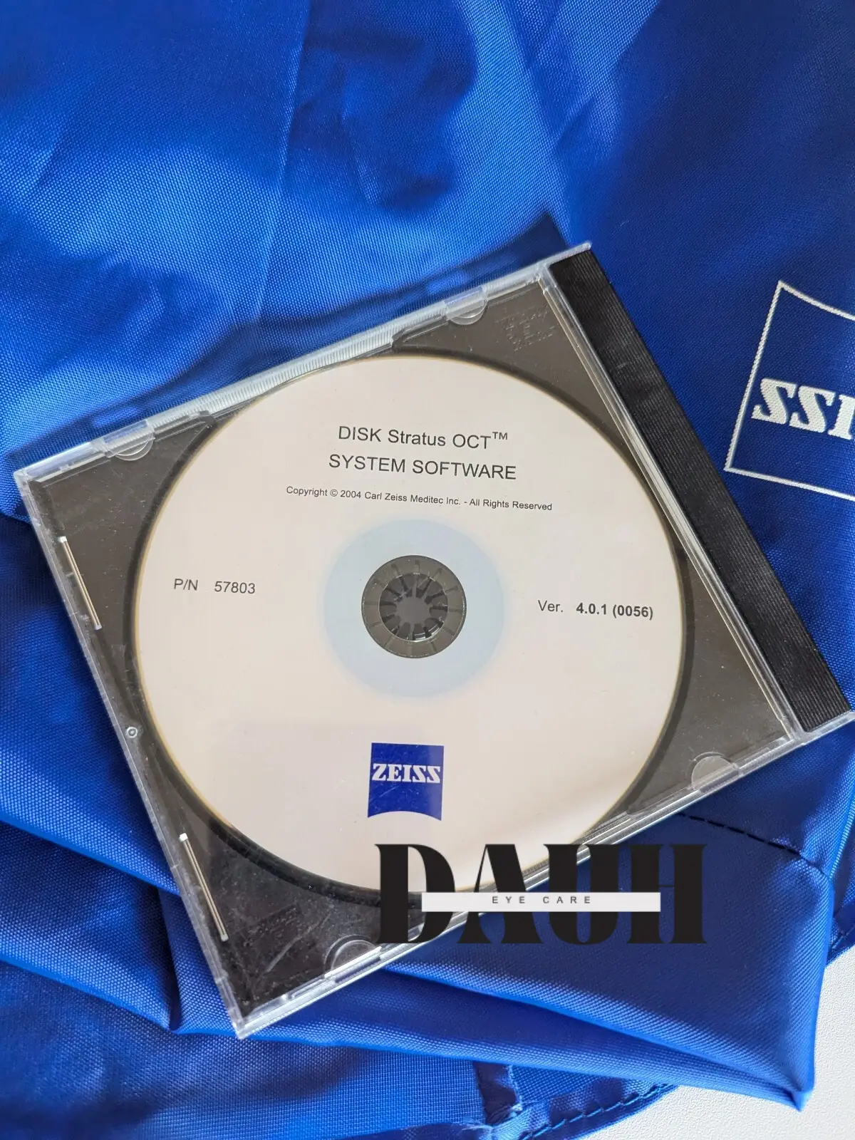
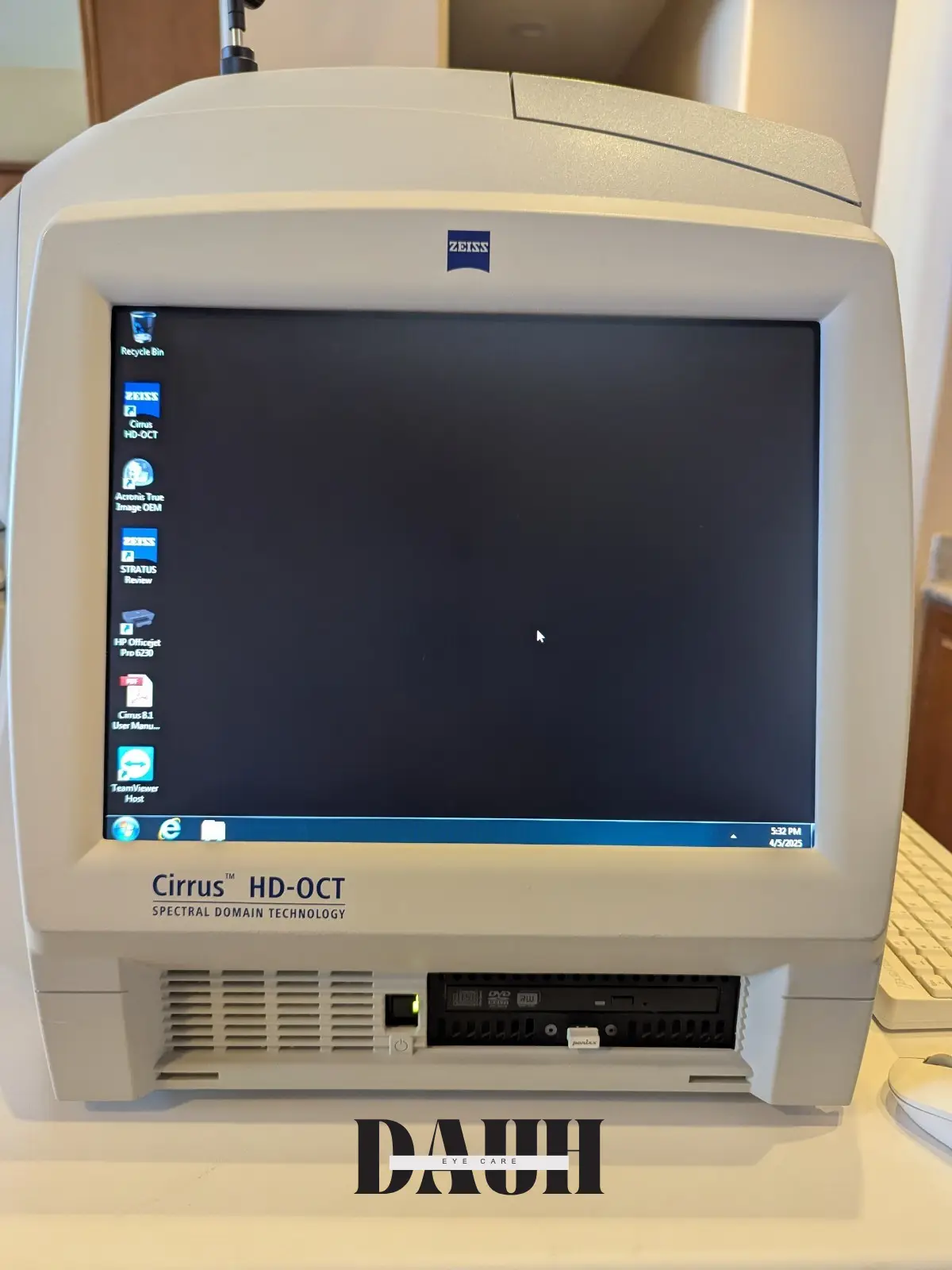
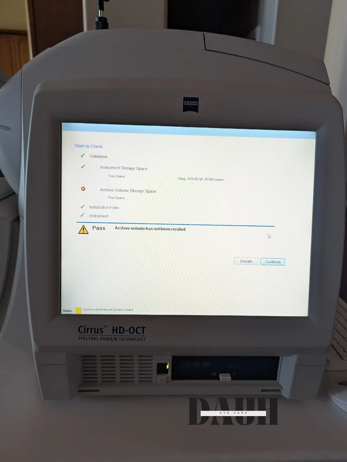

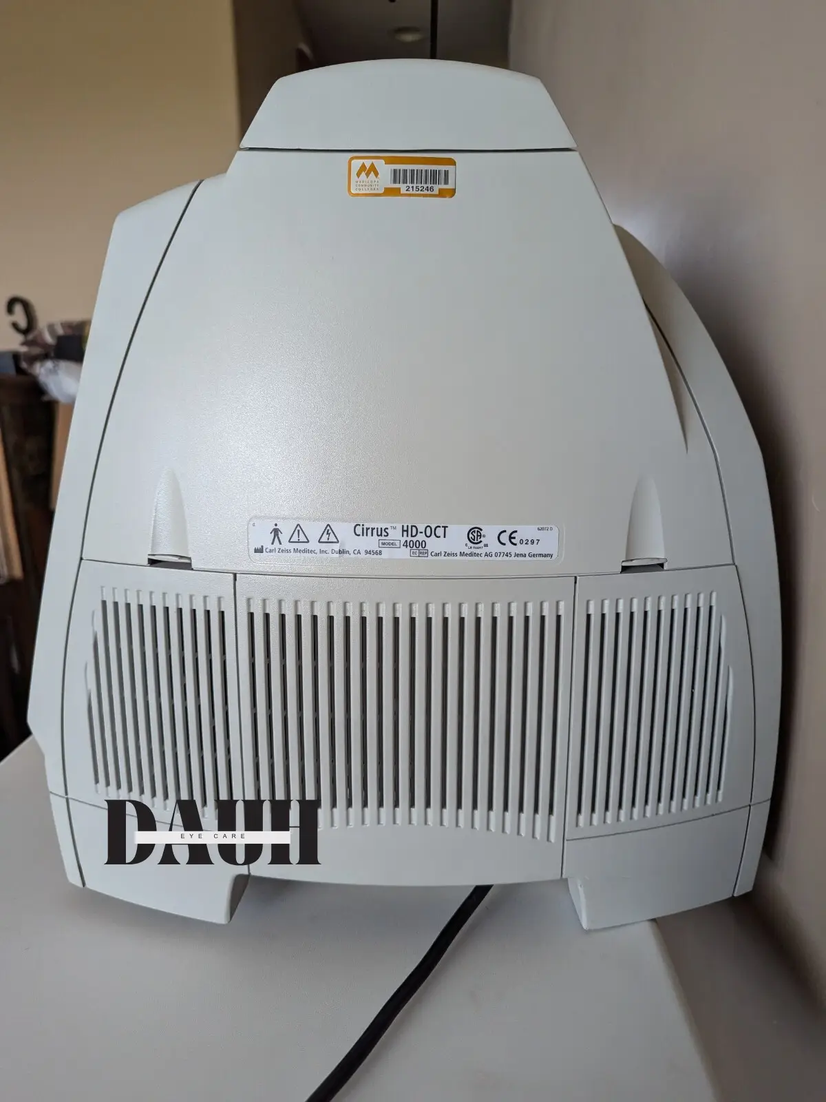
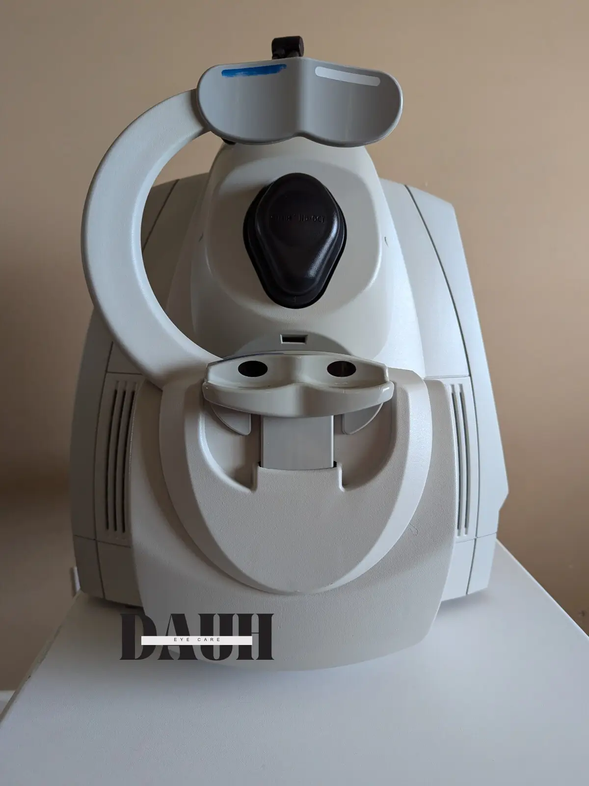
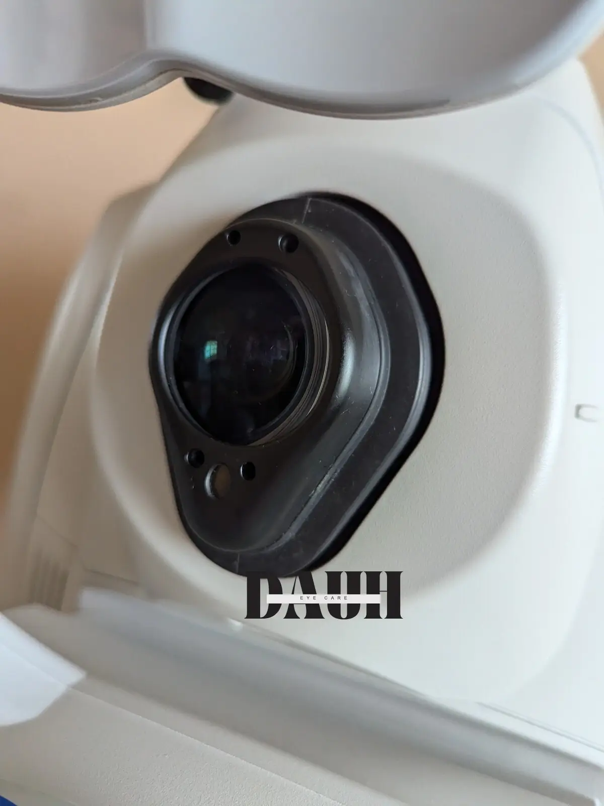
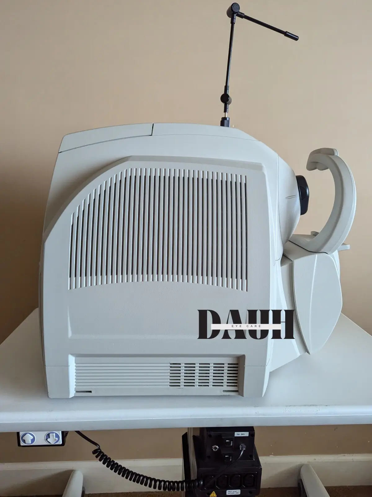
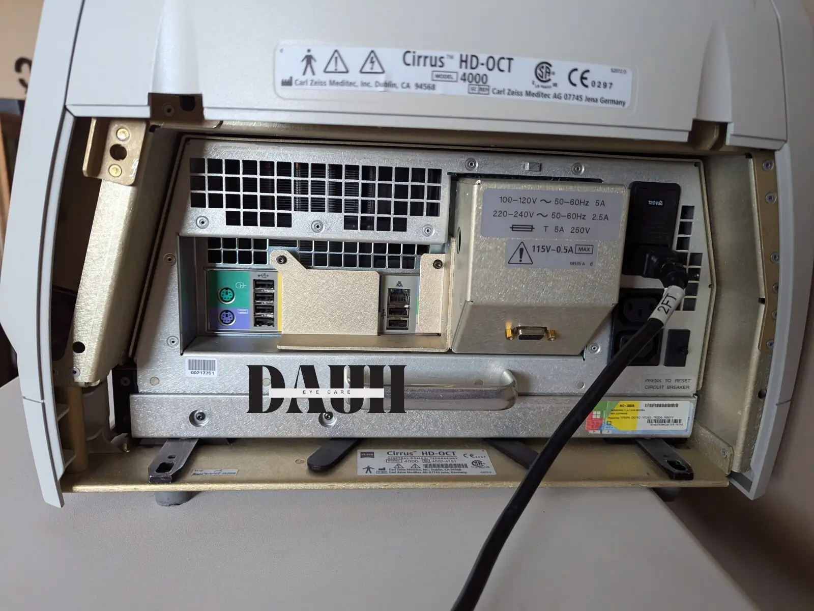
Zeiss Cirrus 4000 HD-OCT
$8,800.00
In stock
Product Description
The ZEISS Cirrus HD-OCT 4000 allows examination of the posterior and anterior of the eye at an extremely fine spatial scale, without surgical biopsy or even any contact with the eye.
Employing the advanced imaging technology of spectral domain optical coherence tomography, Cirrus HD-OCT acquires OCT data about 70 times faster (27,000 vs. 400 A-scans per second) and with better resolution (5 μm vs. ~10 μm axial resolution in tissue), compared to first-generation OCT technology. Cirrus acquires whole cubes of OCT image data, composed of hundreds of line scans, in about the same time as Stratus acquires a six-line scan. You can view these data cubes in three planes, or through three dimensions, giving you access to an extensive amount of retinal image data in one scan.
The Zeiss Cirrus 4000 HD-OCT is a non-contact, high-resolution tomographic and biomicroscopic imaging device. It is indicated for in-vivo viewing, axial cross-sectional, and three-dimensional imaging and measurement of anterior and posterior ocular structures, including cornea, retina, retinal nerve fiber layer, ganglion cell plus inner plexiform layer, macula, and optic nerve head.
The Zeiss Cirrus 4000 HD-OCT normative databases are quantitative tools for comparing retinal nerve fiber layer thickness, macular thickness, ganglion cell plus inner plexiform layer thickness, and optic nerve head measurements to a database of normal subjects.
The Zeiss Cirrus 4000 HD-OCT is intended for use as a diagnostic device to aid in detecting and managing ocular diseases including, but not limited to, macular holes, cystoid macular edema, diabetic retinopathy, age-related macular degeneration, and glaucoma.
Zeiss Cirrus 4000 HD-OCT FEATURES
FastTrac retinal tracking system
Macular Thickness OU AnalysisAdvanced RPE analysis
Ganglion Cell Analysis
Guided Progression Analysis (GPA)
Precision FoveaFinder
Macular Thickness and Change Analysis
Macular Thickness Normative Data
Zeiss Cirrus 4000 HD-OCT SPECIFICATIONS
Technical data
Axial resolution: 5 μm (in tissue)
Transverse resolution: 15 μm (in tissue)
Scan speed: 27,000 A-scans per second
A-scan depth: 2.0 mm (in tissue), 1024 points
Optical source: superluminescent diode (SLD), 840 nm
Fundus Imaging
Line scanning ophthalmoscope (LSO)
Live during scanning
Transverse resolution: 25 μm (in tissue)
Optical source: superluminescent diode (SLD), 750 nm
Field of view: 36° x 30°
Scan Patterns
Macular Cube 200 x 200 Combo: 200 horizontal scan lines comprised of 200 A-scans
Macular Cube 512 x 128 Combo: 128 horizontal scan lines comprised of 512 A-scans
5 Line Raster: 4096 A-scans per B-Scan (adjustable length, spacing and orientation)
HD-OCT Imaging
Methodology: Spectral Domain OCT
Optical Source: superluminescent diode (SLD), 840 nm
Optical Power: < 725 μW at the cornea
Scan Speed: 27,000 A-scans per second
A-Scan depth: 2.0 mm (in tissue), 1024 points
Axial resolution: 5 μm (in tissue)
Transverse resolution: 15 μm (in tissue)
Fundus Imaging
Methodology: Line scanning ophthalmoscope
Live Fundus Image: During alignment and during OCT scan
Optical Source: Superluminescent diode (SLD), 750 nm
Optical Power: < 1.5 mW at the cornea
Field of View: 36 degrees W x 30 degrees H
Frame rate: >20 Hz
Transverse resolution: 25 μm (in tissue)
Iris Imaging
Methodology: CCD Camera
Resolution: 1280 x 1024
Live iris image: During alignment
Zeiss CIRRUS HD-OCT 4000 offers
- Unprecedented visualization of anatomical details
- High-definition layer maps
- High-definition enhanced raster scanning
- Correlations between OCT scans and fundus images
- A 90-degree orientation, which allows the practitioner to observe the patient throughout the exam
- Advanced optics to aid in the examination of patients with cataracts (dilation is not required, even for pupils as small as 2.5 mm)
- Mouse Driven Alignment™, which delivers superior image capture and analysis in just a few clicks, resulting in greater workflow efficiencies
- Auto Patient Recall™, which retains information on the patient’s positioning and instrument settings so they can be repeated for future exams