Optos Monaco Ultra WideField Retina Imaging professionals
$14,000.00
In stock
Optos Monaco Ultra WideField Retina Imaging is the sole UWF imaging device having an integrated OCT to help eye care professionals enhance their clinical examination and enhance practice economics. Imaging modalities and picture viewing options are detailed below.
Product Description
Optos Monaco Ultra WideField Retina Imaging is the sole UWF imaging device having an integrated OCT to help eye care professionals enhance their clinical examination and enhance practice economics. Imaging modalities and picture viewing options are detailed below.
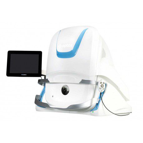
The sole ultra-widefield retinal imaging device with integrated OCT, Optos Monaco Ultra WideField produces a 200° single-capture optomap image in less than 1/2 second and provides cross-sectional 40° OCT perspectives of retinal structures.
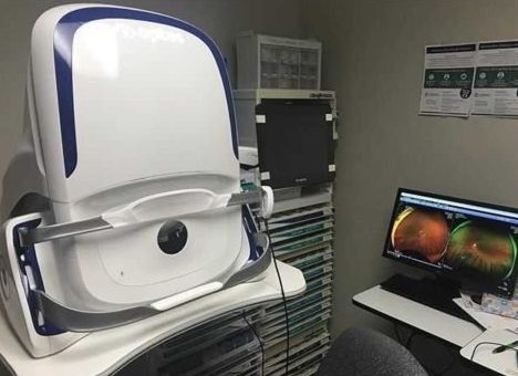
Optos Monaco Ultra WideField is a streamlined desktop UWF retinal imaging apparatus, which provides greater imaging performance, according to a company press release.
The Optos Monaco Ultra WideField is the organization’s first combined imaging device with ultra-widefield color imaging, 3-in-1 color depth imaging and autofluorescence modalities paired using OCT..
Optos Monaco Ultra WideField, long known for its dominance in ultra-widefield imaging, announced today it is bringing spectral-domain OCT capacities to its Monaco desktop imaging device for eye care practices.
In its tomographic imaging manner, Monaco supplies cross-sectional 40° views of retinal structures in 70,000 scans per second with axial resolution beneath 10µm, the company says.
Based on the firm, Optos Monaco is your very first ultra-widefield imager additionally to perform OCT scanning. The accession of OCT”offers a broader collection of imaging tools within 1 machine,” the firm said in a press release,”enabling eye care practitioners to treat and see retinal pathology earlier and more efficiently.”
Image Modalities
- Color
- Red-free
- Choroidal
- Autofluorescence
- OCT
Image Views
- Standard: 200⁰ Single Capture
- Auto-montage: Up to 220⁰
- Central Pole: Detailed view of the macula
- Stereo: Image pairing for optic disc and retinal evaluation
- OCT: Cross-sectional imaging of ocular structures, including the fundus
Monaco offers the following benefits:
• UWF with integrated OCT saves time, space and minimizes patient movement
• Central pole OCT provides comprehensive multi-modal imaging
• optomap images and OCT scans are correlated to facilitate pathology examination
• Color, AF, and OCT images are shown in a single, comprehensive view
Optos Monaco Features and Benefits
• UWF with integrated OCT saves time, space and minimizes patient movement.
• High resolution 200º single-capture optomap images improve pathology detection and management frommacula through the far periphery.
• Non-mydriatic, cSlO imaging through most cataracts and small pupils (2 mm).
• 3-in-1 Color Depth ImagingTM provides important clinical data from the retinal surface through the choroid.
• Green laser autofluorescence minimizes patient exposure to blue light and shows macula and optic nerve head detail.
• Central pole OCT provides comprehensive multi-modal imaging.
• optomap images and OCT scans are correlated to facilitate pathology examination.
• Color, AF, and OCT images are shown in a single, comprehensive view.
• Fast, comfortable image acquisition is easier on patients and improves clinic flow.
• OptosAdvance Image Management facilitates image review and consultations and includes measurement and auto montage capabilities.
Technical Specifications
| Image Modalities | optomap color and optomap plus (red and green laser): Color composite view Green laser view Red laser view optomap af (green laser): autofluorescence Optical Coherence Tomography (OCT) |
|---|---|
| Resolution | optomap color: 20 μm optomap plus, af : 14 μm |
| Wavelengths | Red laser: 635 nm green laser: 532 nm (for af ) |
| Exposure Time | Less than 0.4 seconds |
| Tomographic Imaging | Signal Type: Optical scattering from tissue Signal Source: Super luminescent Diode (SlD) 830 nm Optical Power: laser safety Class-1 following IEC/en60825-1:2014 Typical Axial Resolution: <10 micron (in tissue) Digital on-screen <6 micron Transverse Resolution: 20 micron (in tissue) Scanners: Galavanometric with x, y mirrors Scan Depth: Up to 2.5mm |
| OCT Scan Characteristics | Spectral Domain OCT A-Scan rate up to 70k cycles/s Active eye tracking Automatic scan positioning |
| OCT Scan Types | Line Scan Raster Scan Retina Topography Scan Optic nerve Head (OnH) Topography Scan Retinal nerve Fiber layer (RnFl) Scan |
| Footprint | Width: 550 mm/22 inches Depth: 500 mm/19.5 inches Height: 650 mm/25.5 inches |
https://www.youtube.com/watch?v=qCxN8jMrJxs