SOCT Copernicus REVO with Angio OCT
$8,400.00
In stock
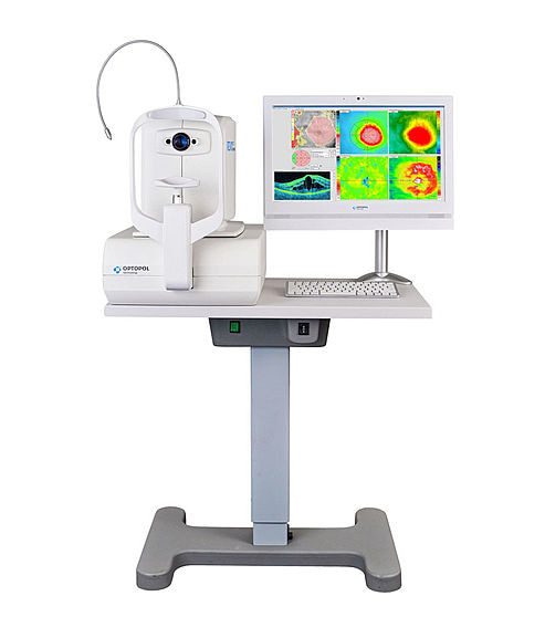
SOCT Copernicus REVO is very easy to use, which allows some personnel freedom. All you need to do is position the patient and start the unit. The automated examination process, which can be carried out even with minimal experience in the OCT domain, features a variety of analysis and review tools. This allows you to use it for either screening or diagnosis, depending on the patient’s needs and condition.
Product Description
SOCT Copernicus REVO is very easy to use, which allows some personnel freedom. All you need to do is position the patient and start the unit.
The automated examination process, which can be carried out even with minimal experience in the OCT domain, features a variety of analysis and review tools. This allows you to use it for either screening or diagnosis, depending on the patient’s needs and condition.OCT made simple as never before.
Position the patient and press the START button to acquire examinations of both eyes. The SOCT Copernicus REVO, using vocal messages, guides the patient through the process of increasing comfort and reducing patient chair time. Creating customized scanning protocols of different diagnostic scenarios will speed up workflow.
This OCT unit features a proprietary spectrometer that will guarantee faster scanning speeds and better quality images across a wider scanning surface. It will help you meet the daily demands of your practice, localize OCT services, and improve the patient experience. The ergonomically designed model also features noise reduction technology for a high definition presentation to improve detection and diagnosis capability.
The SOCT Copernicus REVO is designed for minimal space consumption thanks to the multiple operator and patient positions and reduced system footprint. It can make a patient examination independently, thanks to an automated system that only requires minimal operator interference. The model also offers these features that could help benefit your practice.
Quick scanning
The model features an impressive 80000 A-scan/second speed, which will help improve the examination experience for both the patient and clinician. Little effort and time are required when assessing the eye, with the 3D results produced able to be used for both Retina and Glaucoma analysis.
High detailed imaging
With the non-invasive and dye-free angiography module, you will be able to map out a high definition 3D image of blood circulation in the retina. It also allows you to observe, monitor, and compare any change in the microvasculature in both retinas for a clearer picture. The angiography module features a highly detailed mosaic image that covers a larger field of view of the retina. It presents up to 12 images from different perspectives for more comprehensive service provision.
Comprehensive glaucoma analytics
The model includes specific glaucoma diagnosis and tracking tools. They will help you quantify the ganglion layer, nerve fiber layer, and optic head to increase the precision of your examination and assessment for glaucoma patients. It offers a precise and advanced view, thanks to new optic parameters. You can combine information from both the disc and retina scans to increase the field of view and create a comprehensive picture of glaucoma to help address it better.
Increased productivity
SOCT Copernicus REVO features a comprehensive networking system that is designed to improve the overall patient experience. You can store, transmit, and exchange exam results through a DICOM network. You will be better able to keep track of patient information across different departments for seamless operation. You can also view and manage several examination results through the customized review stations on offer for each practice.
Upgrade potential
SOCT Copernicus REVO also includes two optional additional software modules. Biometry OCT (B-OCT) and Topography OCT (T-OCT) will improve your ability to take high-quality images and compare results across different metrics for multiple visits.SOCT Copernicus REVO offers all the newest standards available in Spectral OCT technology.
COMPREHENSIVE GLAUCOMA SOLUTION
Comprehensive glaucoma analysis tools for quantification of Nerve Fiber Layer, Ganglion layer Optic Nerve Head with DDLS allows for precise diagnosis and the monitoring of glaucoma over time. Asymmetry Analysis of Ganglion layers between hemispheres and between eyes allows the identification and detection of glaucoma in its early stages and non-typical patients. With the golden standard 14 optic nerve parameters and a new Rim to Disc and Rim Absence, the description of the ONH condition is quick and precise.
Invaluable combination of information about the functional quality of vision with comprehensive data on Retinal Ganglion Cells, RNFL, and Optic Nerve Head for both eyes on a single report page. The S&F report contains the following:
- VF sensitivity results (24-2/30-2 or 10-2)
- Total and Pattern Deviation probability graphs for VF results
- Reliability and Global indices for VF results
- Combined map of Structure & Function
- Ganglion cell analysis (GCL+IPL or NFL+GCL+IPL)
- ONH and NFL analysis including charts and comparison tables
- NFL Asymmetry Plot
RETINA
A single 3D macula scan performs both Retina and Glaucoma analysis. The software automatically recognizes eight retinal layers that assist with a precise diagnosis and the mapping of any changes in the patient’s condition.
The SOCT Copernicus REVO 60 000 and 80 000 A-scan/seconds is available now with the Angiography module. This module allows visualization of the retinal microvasculature. Angiography SOCT is a non-invasive, dye-free technique providing a 3D image of retinal blood circulation.
Features:
- Fully automatic. Alignment, focus, and acquisition are fully automated.
- Extremely small dimensions and weights, like an auto refractometer.
- Scans in mioses up to 12mm. Retina and disc are analyzed thanks to a single scan.
- Front segment without any additional lens. It is possible to scan from both iridocorneal angles.
- Connection to any PC thanks to a single USB cable.
- Analysis of 8 retinal layers with the regulatory database.
- Glaucoma analysis with nerve fiber thickness and Gangliari cell analysis with the regulatory database.
- Pachymetric and iridocorneal angle analysis.
- Methods of comparisons and follow-up of exams and pathologies.
- Optional angiography module.