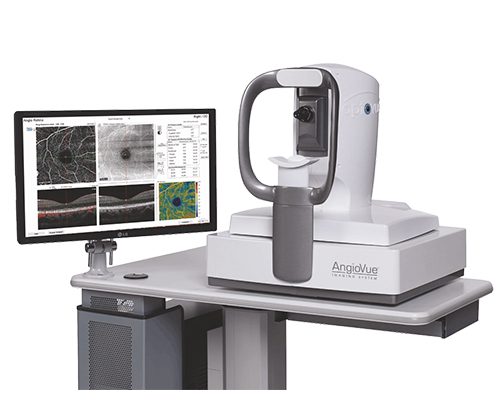
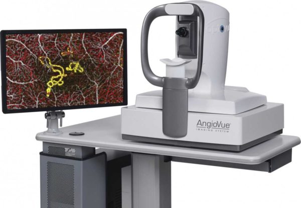

Optovue AngioVue OCT and OCTA Imaging System
$15,000.00
In stock
AngioVue Essential offers high-resolution, noninvasive imaging of retinal vasculature to assist clinicians to visualize ocular disease, the company said in a press release.
Product Description
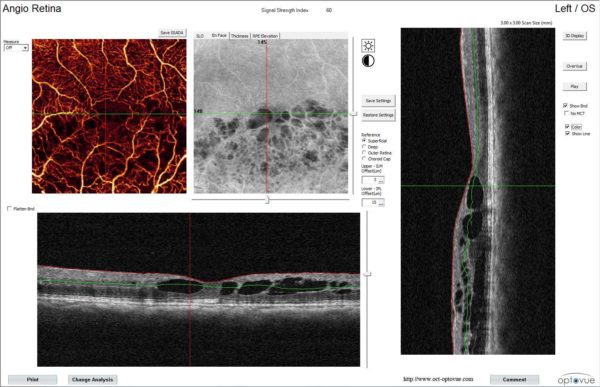
Optovue AngioVue OCT and OCTA Imaging System offers high-resolution, noninvasive imaging of retinal vasculature to assist clinicians to visualize ocular disease, the company said in a press release. The new feature that is OCT-A supplies a report that displays OCT B-scans and layers of a cortical vasculature. The system gives the accession of ocular health screening. It’s designed particularly for practices to permit adoption of OCT-A and OCT to the workflow with minimum disruption.
Optovue AngioVue OCT and OCTA Imaging System provides retinal experts with the ability to quickly visualize the existence or absence of retinal vessels and assess new information concerning the microvasculature in detail.
The info may be integrated with other diagnostic imaging results to form a complete picture of a patient’s disease condition and their treatment choices.
By adding AngioVue Retina into our product portfolio, we can help retinal experts acquire OCT-A technology in a cost-efficient manner, thereby adding this technology to their armamentarium of diagnostic imaging applications.” A noninvasive imaging program, it could imagine microvascular blood circulation in less than three seconds. It is intended to help clinicians make therapy choices by analyzing structure and purpose. These imaging capabilities make it easier for physicians to customize patient care and follow treatment and disease progression in detail.
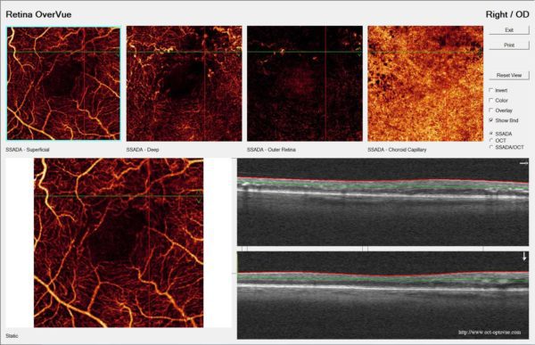
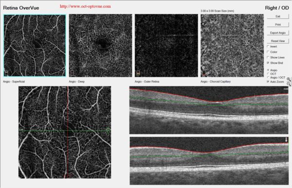
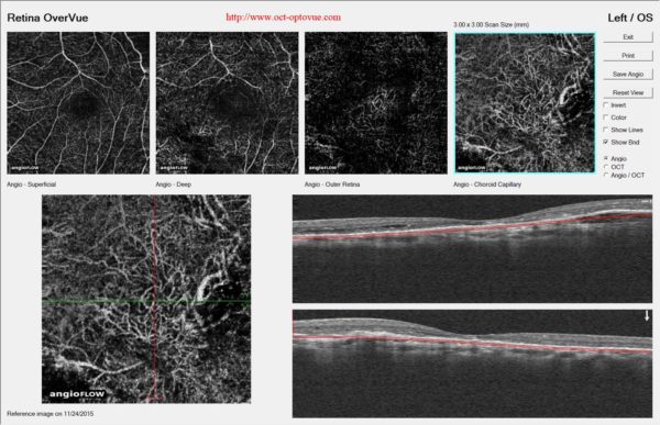
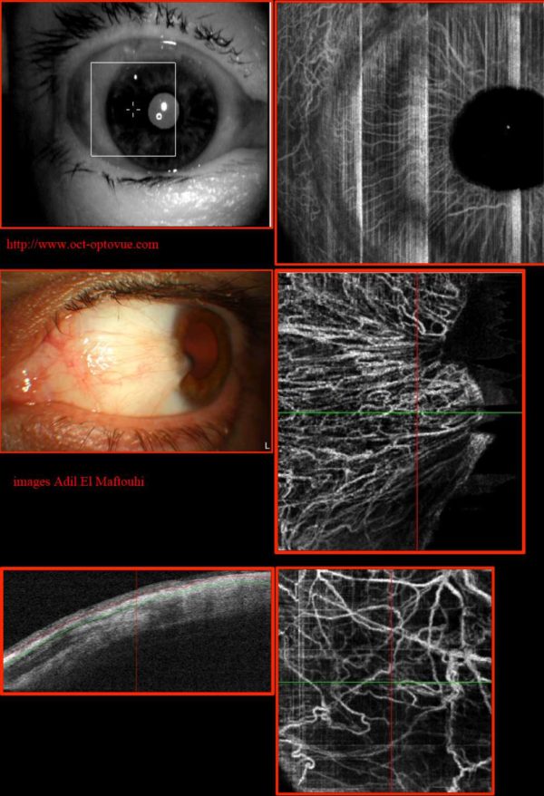
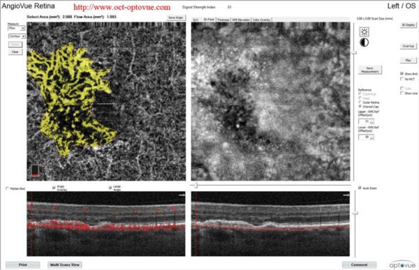
Optovue AngioVue OCT and OCTA Imaging System makes it possible for clinicians to image the motion of scattering particles like erythrocytes (red blood cells) using sequential OCT cross-sectional scans that are replicated in the same location on the retina. Capturing the dynamic movement of the erythrocytes (red blood cells) allows a 3D visualization of the perfused vasculature and microvasculature of the retina. Unlike Angiovue permits visualization of vasculature within specific layers of the retina, without the effects of pooling or discoloration.
Optovue AngioVue OCT and OCTA Imaging System Key Features
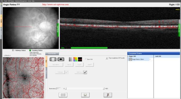
- Fast, Non-Invasive Imaging Quickly visualizes your patient’s microvasculature with a non-invasive, dye-free technique that can be accomplished by your technician in a matter of seconds. With AngioVue, you can image your patients as often as needed to closely follow disease progression and treatment response.
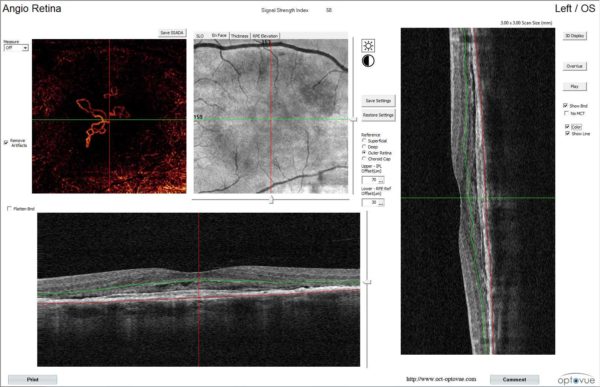
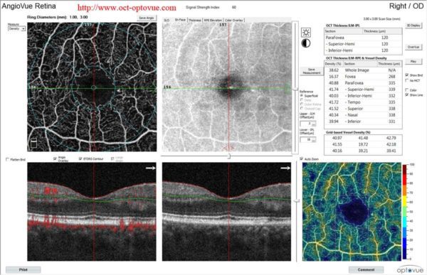
- Designed for Visualization Even in the Choroid AngioVue’s Outer Retinal Zone is a single en face slab that visualizes a wide spectrum of pathologies arising from abnormal vessel growth in the retinal-choroidal interface, such as Type 1 and 2 CNV. The AngioVue Imaging System utilizes a proprietary projection artifact removal technique resulting in unparalleled image quality uniquely suited for enhanced neovascularization visualization.
- Proven Performance Trust the system that has been installed in more than 550 practice worldwide and featured in more than 90 peer-reviewed publications. Partner with a company committed to industry-leading clinical education, technical support and product innovation.
- Exclusive Technology The AngioVue Imaging System uses the proprietary SSADA algorithm to produce stunningly detailed images and to minimize scan acquisition time. Optovue’s exclusive Motion Correction Technology (MCT™) reduces motion artifact to give you detailed images that increase your diagnostic confidence.
The combination of five important components:
- Fast spectral-domain OCT (70,000 A-scans/sec)
- Patented Motion Correction Technology (MCT)
- Patented Split Spectrum Amplitude Decorrelation Angiography (SSADA)
- CUDA parallel processing architecture
- Patented enface visualization of 3D OCT data
Specifications:
- OCT Camera : 70,000 A-scans per second
- Optical Resolution: (in the structure)
- Depth: 5.0μm
- Beam Spot Size: 22μm
Image Sampling Rate:
- Depth: 3.0μm digital resolution
Scan Range:
- Depth: ~3mm
- Transverse: 2mm to 12mm
- Scan Beam Wavelength: =840±10nm
- Exposure Power at pupil: 750μW maximum
Patient Interface:
- Working Distance: 22mm
- Motorized Focus Range: -15D to +20D