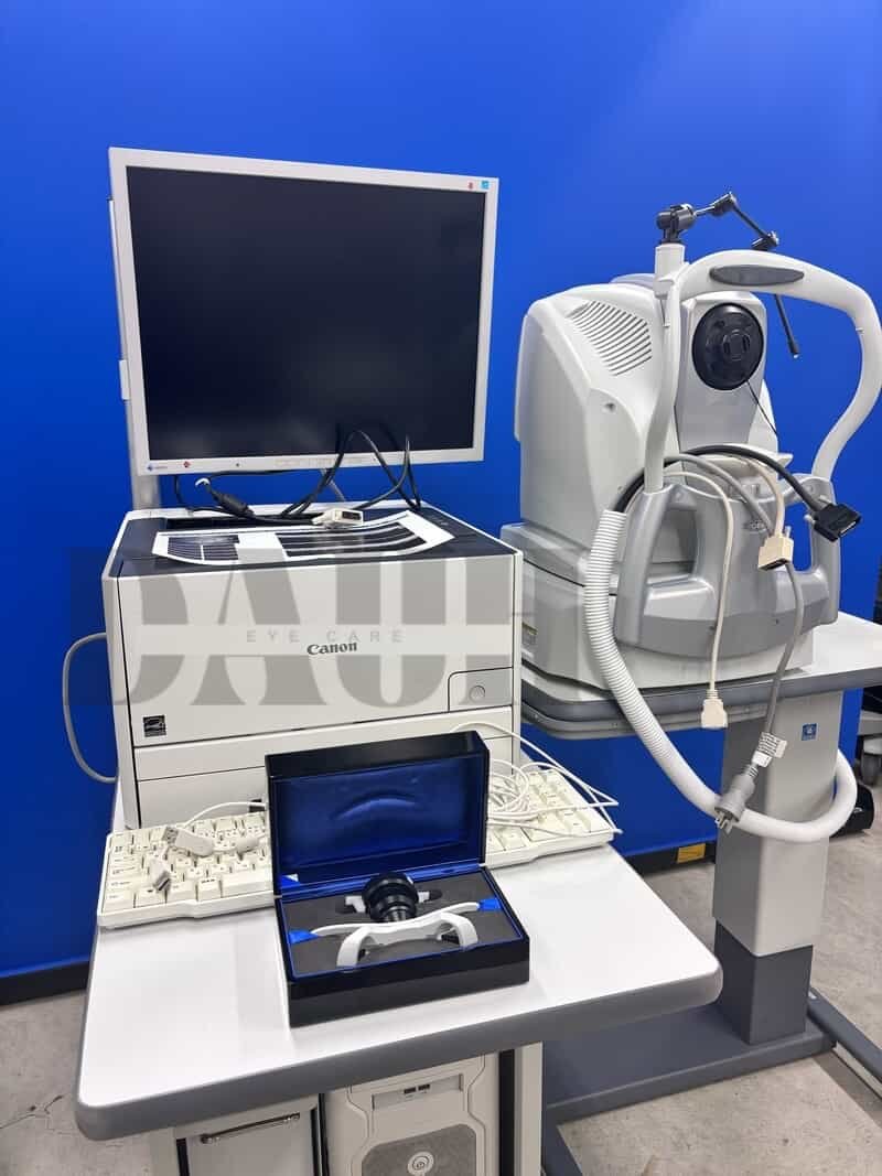

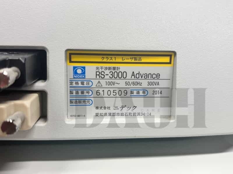
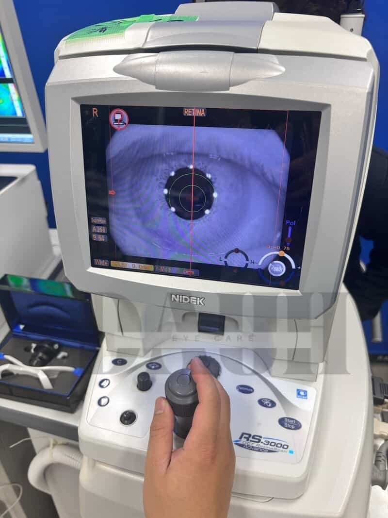
NIDEK RS-3000 Advance OCT system
$8,900.00
In stock
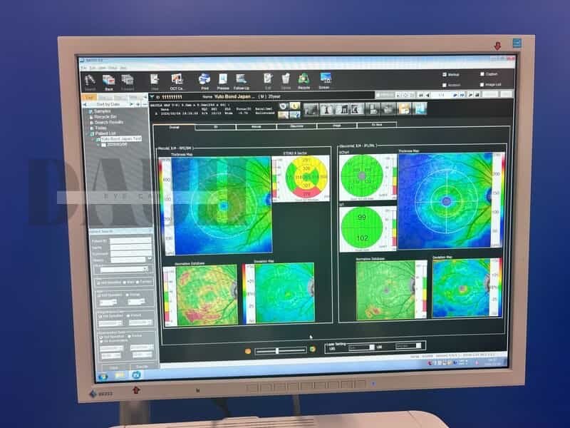
NIDEK RS-3000 Advance is a premier OCT system incorporating Scanning Laser Ophthalmoscope that is designed for comprehensive evaluation of the retina and choroid. The NIDEK RS-3000 Advance OCT provides exquisite detail of the retinal and choroidal microstructures to assist in clinical diagnosis
Product Description
NIDEK RS-3000 Advance OCT is a premier OCT system incorporating a Scanning Laser Ophthalmoscope that is designed for comprehensive evaluation of the retina and choroid.
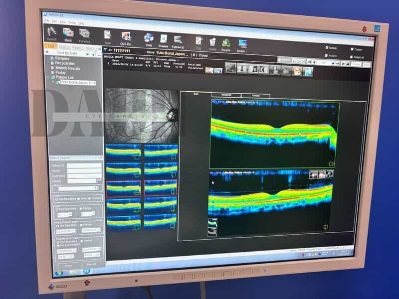
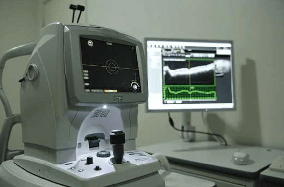
The NIDEK RS-3000 provides exquisite detail of the retinal and choroidal microstructures to assist in clinical diagnosis. The combination of Retinal Camera and OCT saves space and provides comprehensive information for clinical diagnosis.
Nidek has announced the launch of the RS-3000 Advance 2 optical coherence tomography (OCT) system, which incorporates a scanning laser ophthalmoscope (SLO) and is designed for comprehensive imaging and analysis of the retina and glaucoma.
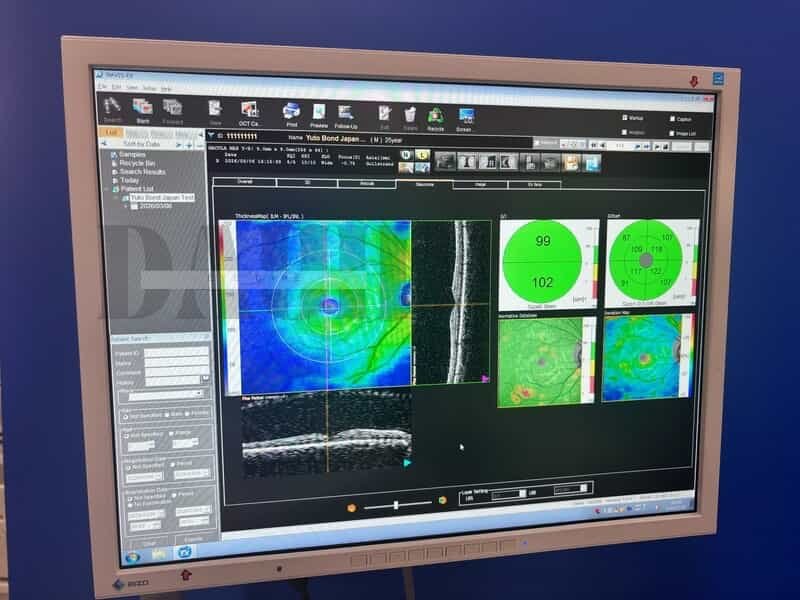
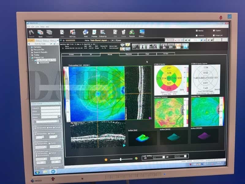
NIDEK RS-3000 Advance OCT retains some of the signature features of the previous model, including glaucoma analysis with a 9 x 9 mm wide area normative database, accurate image capture with an SLO-based eye tracer, and selectable OCT sensitivity that allows acquisition of B-scan images through media opacities. Enhancements of the RS-3000 Advance 2 include 85,000 A-scans/s, 12 x 12 mm auto-panorama imaging for OCT angiography, and improved image quality.
The faster scanning speed allows rapid image acquisition, decreasing patient chair time and increasing clinical efficiency. Higher scanning speeds also reduce artifacts due to fixation loss, resulting in better image quality. The 12 x 12 mm panoramic function enables imaging of peripheral lesions resulting from retinal pathology in branch retinal vein occlusion and diabetic retinopathy.
The tracing HD plus function traces involuntary eye movements to maintain the same scan location on the SLO image for accurate image capture. This function allows accurate averaging of up to 120 images.
Key Features
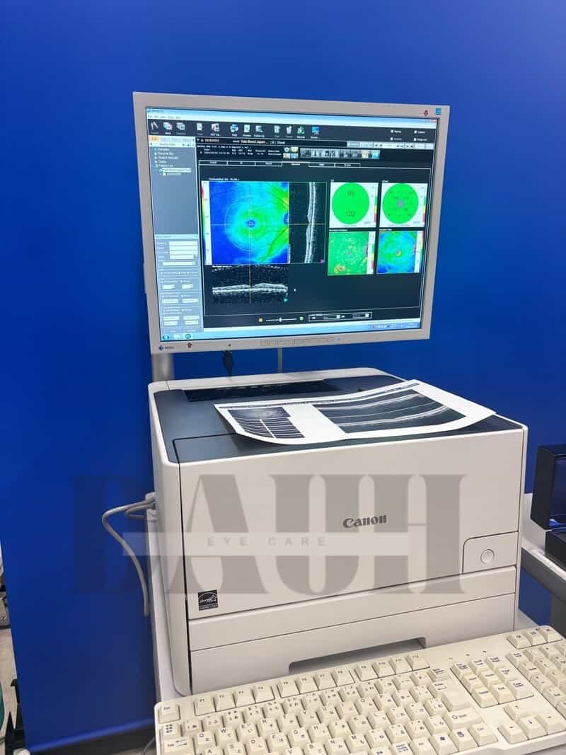
- Retinal Camera and OCT combined in one instrument
- Anterior imaging for pachymetry
- Anterior imaging for angle view
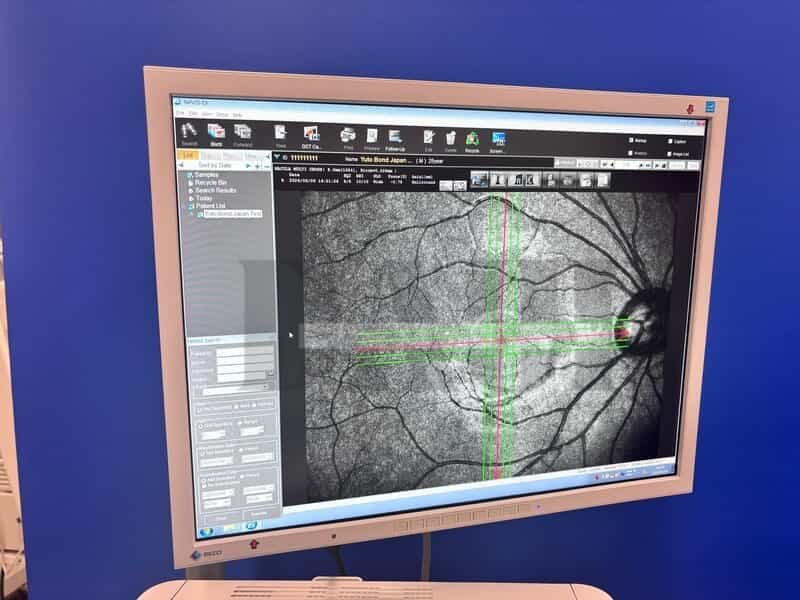
- Superb macula image using SLO technology
- Wide-field imaging, from macula to disc
- Tracing HD plus for accurate averaging of maximum 120 images
- High speed scanning at 53,000 Ascans/sec
- Selectable OCT sensitivity based on ocular pathology enables enhanced visualization
- Torsion Eye Tracer (TET) for accurate image capture
- Multifunctional follow-up Customized report
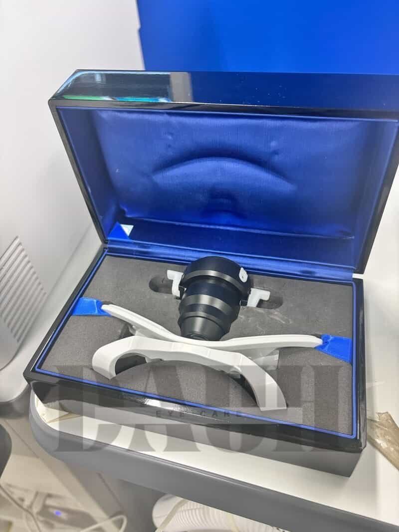
Include: Main OCT Unit, a keyboard, PC, Monitor