Combining comprehensive aspects of Topcon Swept Source, Topcon guarantees high-quality imaging. It takes into account different aspects of imaging to guarantee high-resolution fundus imaging, which will ensure better quality images for a more accurate assessment. These are some features of the DRI OCT Triton that should make it ideal for your practice.
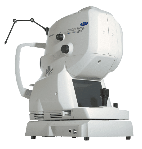
Topcon Swept Source DRI OCT Triton is a multi-modal swept-source OCT with a non-mydriatic color fundus camera. Utilizing a 1,050 nm wavelength light source, and a scanning speed of 100,000 A Scans/sec, it provides uniform scanning sensitivity allowing superior visualization of the vitreous and choroid in the same scan.
Invisible OCT scanning light, eye tracking during the capture of selected scans, along with high scanning speeds reduce the effect of patient eye movement, improving workflow and allowing for more data be to collected in a shorter period of time. A 12 mm x 9 mm widefield scan along with automated layer segmentation provides measurement and topographical maps, with reference database, of the optic nerve and macula in one scan.
The Triton Plus model also includes a monochrome camera for fluorescein angiography and fundus autofluorescence utilizing the exclusive Spaide autofluorescence filters.
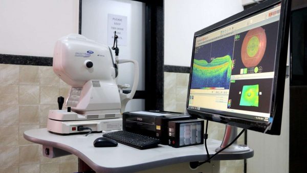
The easy-to-use, intuitive IMAGEnet 6 software enables dynamic viewing of the OCT data, providing 3D, 2D and fundus images simultaneously. Pin-Point™ Registration identifies an exact pathological location across all imaging modalities available within the Triton. In addition, both compare and trend analysis functions allow users to view serial exams of several scan protocols including our 12x9mm 3D Widefield scan. En face technology*, with layer flattening application allows for visualization of the various layers of the retina.
Increased penetration
At 100,000A-Scans per second, which is roughly double the average scan speed, this OCT will offer better output than its competitors at an efficient rate. It features SWEPT source OCT, which is designed for better visualization in a single scan. It also features a larger and deeper scan surface for increased detail.
Automatic tracking
The model features automatic scanning and tracking that facilitates accurate imaging and monitoring of patients’ conditions. It automatically places any subsequent scans on specific positions to allow accurate monitoring of changes made from the initial scan results. This will enable the better assessment and facilitate higher quality services for your patients. Active eye tracking will help map the eye, track any movement and account for the original structure to ensure that any blinks and sudden ocular movements do not compromise the accuracy of your findings.
Anterior segment analysis extension
The model’s SWEPT source technology can be extended to include anterior imaging, which facilities better diagnosis. This attachment will increase the sharpness of images, allowing for higher accuracy during imaging. It will work well even for peripheral parts of the examination, such as the corneal edges and in-depth imaging of the anterior chamber. Your clinicians will receive high-quality imaging of even naturally difficult parts. This will allow for more accurate assessment and diagnosis.
Improved fundus imaging
The low-intensity flash of this model utilizes laser lighting as opposed to halogen lighting. The result is a clearer image with less likelihood of a glare. The high resolution and high contrast image offered by the DRI OCT Triton will feature true color and do away with noise.
This will help you get the most accurate readings every time. It increases the diagnostic options available to your practice with additional imaging modules such as multi-color fundus imaging, fundus autofluorescence and fluorescent angiography for a wide range of imaging options. The better resolution imaging will boost the performance of your practice in terms of high-quality output and services offered.
Efficient scanning
The Topcon Swept Source features invisible scan lines, which reduces the likelihood of patient distraction. OCT models with visible scan lines will often require re-scanning because patients may be more likely to blink and engage in other eye movements as a result of the distraction from these lines. It will improve repeatability and reduce the amount of time required to get sufficient information.
The DRI OCT Triton also combines multiple images of the disc and macular areas into a single shot, which offers clarity and detail without requiring too much effort. During the assessment, your clinician will be able to carry out macular and RNFL assessment without using multiple images. Their diagnosis will be more likely accurate and convenient for use in coming up with treatment suggestions.
Welcome to the New Frontier in OCT Imaging
Swept Source technology & 1,050nm wavelength
Topcon Swept Source provides a significant improvement over conventional OCT. Due to the optimized long wavelength scanning light (1,050nm), there is better penetration of the deeper layers of the eye. Furthermore, this scanning light also penetrates better through cataracts, hemorrhages, blood vessels, and sclera.
The world’s fastest scanning speed 100,000 A-scans/second
Approximately twice higher scan speed, compared to Topcon SD-OCT, will bring more scans for a single B-scan image, and a more informative image supports efficiency and quality of diagnosis.
Better penetration
The high penetration of the Swept Source light can easily and clearly visualize deep layers in the eye, such as choroid and sclera. A further benefit of Swept Source is that it can clearly visualize both the vitreous and choroid in a single scan, that are uniformly clear and noise-free. This eliminates the need for time-consuming vitreous/choroidal combination scans.
Wide and deep scans
In one single image, the vitreous & choroid are revealed in a crystal clear way. The Topcon DRI OCT Triton enhances visualization of outer retinal structures and deep pathologies. The Topcon DRI OCT Triton automatically detects 7 boundaries including the chorio-scleral interface. The 12mm B-scan covers both the macular area and the optic disc.
Invisible scan lines
The invisible 1,050nm wavelength does not distract patients. Patients do not see the scanning line, which is an advantage with elderly patients and children. Reduction in movement artifacts and increased repeatability.
Time efficiency – create one single overview
Combination scans cover the macular and disc areas in a single shot and offer both macular and Retinal Nerve Fiber Layer (RFNL) analysis. Combination scans are time efficient for the operator and convenient for the patient. Combination scans allow both macular and disc analysis in one overview.
Multimodal fundus imaging
The Topcon Swept Source offers a true color, non-mydriatic fundus image while using a very low-intensity flash. This unique feature is a perfect tool for identifying the location of scans in the eye utilizing TOPCON’s patented Pinpoint RegistrationTM. The DRI OCT Triton Plus offers a wide range of diagnostic options with multi-modal color fundus imaging, Fluorescein Angiography (FA) and Fundus Autofluorescence (FAF) for even more diagnostic possibilities. For the first time, Pinpoint registration will be available with fundus autofluorescence and Swept Source OCT.
New tracking system – SMARTTrack
SMARTTrack is a very useful tool to compensate for the ever-present involuntary eye movements (microsaccades). It allows the automatic acquisition of a follow-up scan in precisely the same anatomical location. SMARTTrackTM enhances the user-friendliness of the machine.
Anterior segment analysis
The Topcon DRI OCT Triton can be extended to include anterior imaging, making the Swept Source a versatile diagnosis tool for both anterior and posterior imaging. The anterior attachment ensures sharp images, even in the periphery of the cornea and in-depth images of the anterior chamber.